近期,必发集团welcome欢迎光临汤晓智教授、冯潇副教授联合中国农业大学李媛教授在国际化学类一区期刊Journal of Colloid And Interface Science (Q1, IF=9.7)发表题目为“Germination-induced nanoarchitectonic assembly of quinoa protein at neutral pH and its aerogels for microplastic removal”的研究性论文。
成果亮点:
1. 首次实现中性pH条件下藜麦蛋白的纤维化,无需强酸处理。
2. 揭示了萌发促进藜麦蛋白纤维化的机制,验证了藜麦蛋白纤维良好的生物安全性。
3. 萌发8小时效果最佳,过长的萌发(16、24小时)反而导致无定形聚集,降低纤维化效果。
4. 菠萝蛋白酶水解和二硫键异构酶还原协同处理藜麦11S球蛋白可显著提升纤维转化率。
5. 气凝胶对水体和饮料中的微塑料表现出高效的吸附去除性能。
研究背景:
微塑料已成为全球关注的新型环境污染物,广泛存在于大气、土壤和水体中。研究表明,微塑料可诱导氧化应激、干扰代谢、破坏肠道微生物平衡、影响心肺和免疫系统,并对生殖健康产生负面影响。随着塑料制品的广泛使用,微塑料的全球排放量预计将从2000年的41千吨增长至2060年的1397千吨,形势严峻。目前,研究者已开发出多种气凝胶材料用于去除水中微塑料,如聚多巴胺/壳聚糖气凝胶、还原氧化石墨烯气凝胶、蛋清蛋白气凝胶等。然而,这些材料大多成本高、来源不可持续,或制备过程复杂。植物蛋白因其成本低、可降解、来源广泛等优势,成为理想替代材料。然而,植物蛋白通常凝胶性较差,难以形成稳定的多孔结构,限制了其在气凝胶领域的应用。
研究表明,植物蛋白在强酸条件下加热可发生水解并形成淀粉样纤维,从而改善凝胶性能。然而,这种方法能耗高、对设备腐蚀严重,不利于工业化推广。因此,亟需发展一种温和、绿色、高效的蛋白纤维化诱导方法。萌发是一种天然、低成本的生物改性方法,可激活种子内源酶系,部分水解储藏蛋白,改变其结构和功能特性。藜麦作为一种天然耐逆作物,萌发迅速,适合作为研究对象。在本研究中,首次发现并报道了通过萌发修饰蛋白构象诱导藜麦蛋白在中性pH下纤维化的现象。同时,全面研究了萌发对藜麦蛋白组分、理化性质、构象的影响,以解释其纤维化机制。此外,还探讨了萌发对凝胶微观结构和流变性质的影响。此外,首次采用协同酶法诱导藜麦11S球蛋白纤维化,以提高纤维转化率(42.7%),并生成了功能性气凝胶用于去除水和饮料中的微塑料。本研究为通过绿色策略构建具有潜在微塑料去除效能的藜麦蛋白气凝胶提供了理论基础,并为体外微塑料去除提供了新思路。
主要内容:
本研究首次报道了一种通过种子萌发修饰藜麦蛋白结构促进其在中性pH条件下形成淀粉样纤维的绿色策略,并基于此构建了高效去除微塑料的复合气凝胶。研究发现,藜麦萌发8小时提取的分离蛋白在95 ℃加热30分钟即可形成200–800 nm的纤维结构,其机制在于萌发激活了内源蛋白酶和二硫键异构酶,适度水解蛋白的同时还原二硫键、增强蛋白表面电负性(-30.6 mV),从而通过解折叠蛋白结构、增强静电排斥促进蛋白分子有序排列和自组装;而过长萌发(16、24小时)则因电负性下降和二硫键重新形成导致无定形聚集体增多。本文揭示了2S清蛋白不具备纤维化的能力,而11S球蛋白是形成淀粉样蛋白纤维的主要成分。受萌发机制启发,进一步采用菠萝蛋白酶水解联合二硫键异构酶还原协同处理藜麦11S球蛋白,在中性条件下实现了高达42.7%的纤维转化率,所得纤维长度达2.9 μm,β-折叠含量升至54.2%,且纤维由2–3条原纤维螺旋缠绕而成。进一步,将所得蛋白纤维与壳聚糖复合制备气凝胶(质量比3:1),不仅具有高比表面积和密集网络结构,对水中聚苯乙烯微塑料的吸附容量达54.4 mg/g。在实际饮料体系中,该气凝胶对可乐、雪碧和橙汁中微塑料的吸附容量分别达286.7、457.5和370.9 mg/g。此外,蛋白纤维聚集体在模拟胃肠消化后仍部分保留纤维结构,且对肠上皮细胞无毒性甚至可促进增殖,证明其具有良好的生物安全性。总之,本研究有助于藜麦蛋白的绿色加工,对于植物性蛋白产业和微塑料去除具有重要意义。
图文赏析:
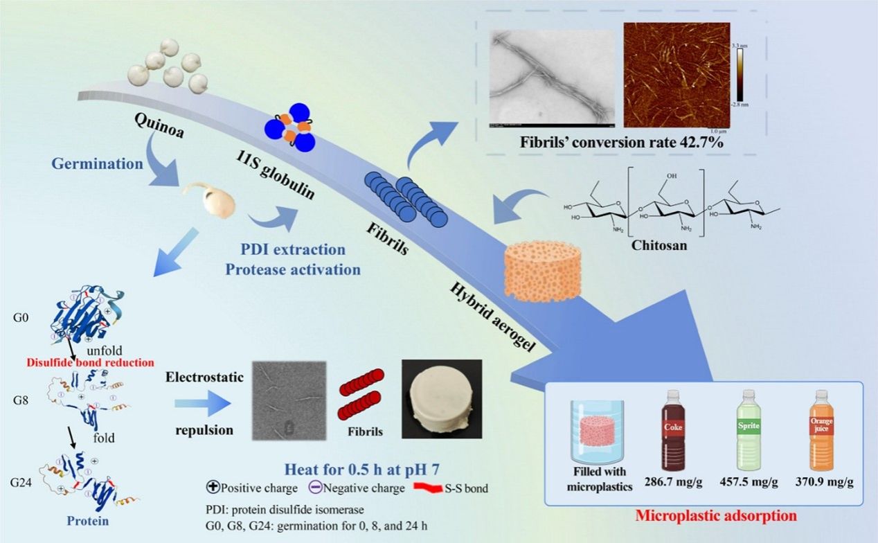
Fig. 1. Graphical abstract
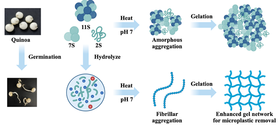
Fig. 2. Hypothesis of this research
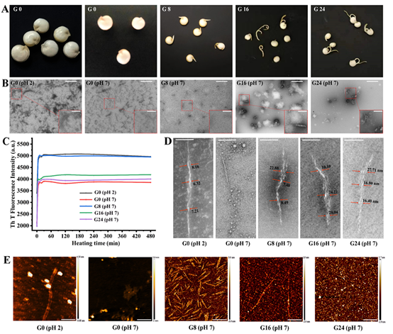
Fig. 3. The effect of germination on the morphology of QPI aggregates and their fibrillation kinetics. (A) Appearance of germinated quinoa. (B) TEM images of QPI aggregates, scale bars, 1 m. Inset zoom image, scale bars, 500 nm. (C) Fibrillation kinetics of QPI. (D) The morphology of QPI fibrils, scale bars, 100 nm. (E) AFM images of QPI aggregates, scale bars, 400 nm. *G0, G8, G16, and G24 indicate QPIs extracted from quinoa germinated for 0, 8, 16, and 24 h, respectively
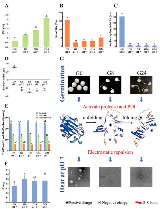
Fig. 4. Effects of germination on the physicochemical properties of QPIs. (A) Degree of hydrolysis. (B) Protein solubility. (C) Surface hydrophobicity. (D) Zeta potential. (E) Total/free sulfhydryl and disulfide bond content. (F) Disulfide bond reduction activity of protein disulfide isomerase extracted from quinoa germinated for 0, 8, 16, 24 h. (G) The mechanism of QPI fibrillation facilitated by germination. *Different lowercase and uppercase letters indicate significant differences (P < 0.05). In Fig. 3D, the significant difference of the absolute value of zeta-potential was evaluated. G0, G8, G16, and G24 indicate QPIs extracted from quinoa germinated for 0, 8, 16, and 24 h, respectively
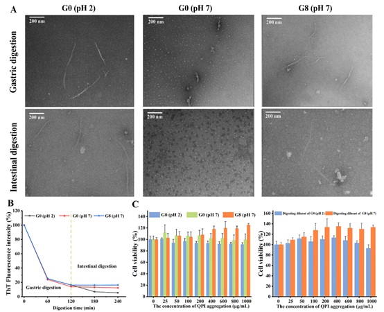
Fig. 5. Effects of gastrointestinal digestion on QPI aggregates. (A) The morphology of QPI aggregates after gastrointestinal (2 h) and intestinal digestion (2 h). (B) Normalized ThT fluorescence intensity of QPI aggregates as a function of digestion time. (C) Effects of gastrointestinal digestion on the cytotoxicity of QPI aggregates. *G0 and G8 indicate aggregates generated from QPI extracted from quinoa germinated for 0 and 8 h, respectively
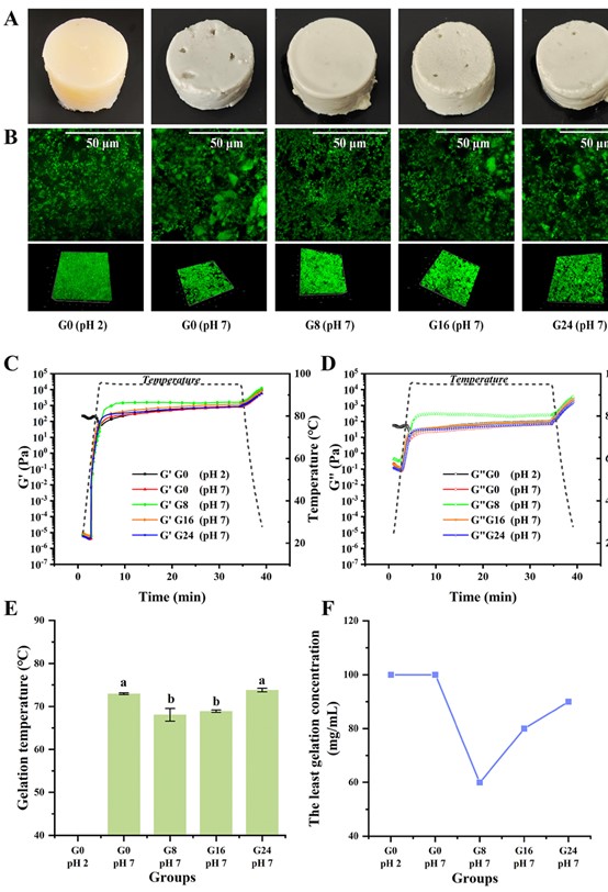
Fig. 6. The effects of germination on the gelling properties of QPIs. (A) Appearance and microstructure (B) of QPI gels. The rheological properties (C, D), the gelation temperature (E), and the lowest gelation concentration (F) of QPI dispersions
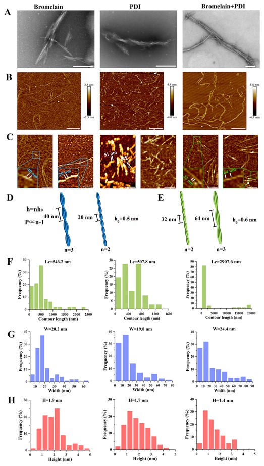
Fig. 7. TEM, AFM and statistical analysis of quinoa 11S globulin fibrillar aggregates with various enzyme treatments. (A) TEM images, scale bars, 200 nm. (B) AFM images, scale bars, 1 m. (C) Magnified AFM images, scale bars, 300 nm. Schematic illustration of the helical fibrils induced by bromelain (D) and bromelain + PDI (E) treatment. (F) Contour length distribution. (G) Width distribution. (H) Height distribution. *h represents the maximum height of the fibril, and h 0 resents the height of the protofibril. n represents the number of protofibrils, and P represents the periodic pitch of fibril
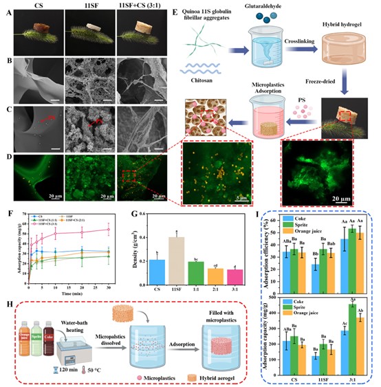
Fig. 8. Characterization and microplastics adsorption of aerogels. (A) Photographs of light aerogels on top of setaria viridis. (B, C) SEM images of aerogels before and after adsorption of microplastics. Scale bar, 20 μ m. (D) CLSM images of aerogels after adsorption of microplastics. Scale bar, 20 μ m. (E) Schematic illustration of the synthesis process and microplastics adsorption of hybrid aerogel. (F) Microplastics adsorption capacity versus time at an initial concentration of 100 ppm. (G) Density of aerogels. (H) Schematic illustration of the microplastics adsorption of hybrid aerogel in three beverages. (I) The effect of aerogels on the adsorption efficiency and adsorption capacity of microplastics in different beverages. *Letters a-c and A-C in Fig. 7I denote statistically significant differences in microplastic adsorption among different beverages by the same type of aerogel, and within the same beverage by different types of aerogels, respectively
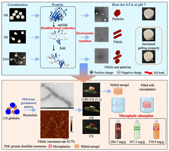
Fig. 9. Mechanistic illustration of germination-induced fibril nanoarchitectonics of quinoa protein at neutral pH and its aerogel application for microplastic removal
原文链接:https://doi.org/10.1016/j.jcis.2025.139058